Clubfoot vs. PreClinical Clubfoot Deformity. A Technical Presentation
The first published paper on The PreClinical Clubfoot Deformity was in 2002 (Rothbart BA, 2002). Below is an embryological discussion on the differences between the Clubfoot and PreClinical Clubfoot Deformity.
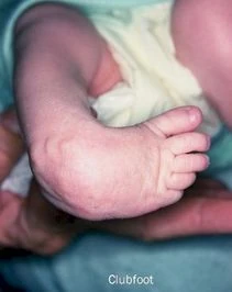
Clubfoot Deformity
During the embryological development of the foot:
- the calcaneus fails to unwind (remains in relative supinatus)
- the cuboid remains in supinatus, and with it, the lateral two cuneiforms, lesser four metatarsals and accompanying phalanges (Bohm,1929)
Since the footplate always unwinds heel to toe (Streeter, 1945, 1948, 1951), as goes the calcaneus, so goes the talus.
Retention of calcaneal supinatus is always accompanied by talar supinatus, the Clubfoot deformity (Bohm 1929). In the postnatal (after birth) foot, the calcaneus articulates with the cuboid, which in turn articulates with the navicular, lateral cuneiform and lateral two metatarsals.
From a structuralist viewpoint, one might conclude that the calcaneus only impacts these structures. From an embryological prospective, this is not the case. Bohm (1929) describes how the ontogenetic unwinding of the calcaneus affects the lateral column of the embryonic foot. That is, the relative structural position of the cuboid, the lateral two cuneiforms and four lateral metatarsals/phalanges are determined by the sculpturing of the calcaneus.
Retention of calcaneal supinatus in the prenatal foot is manifested as the Clubfoot Deformity in the postnatal foot (See photo below).
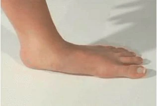
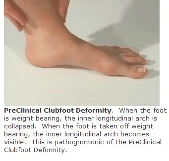
PreClinical Clubfoot Deformity
If the calcaneus has unwound enough, that is, where the cuboid is no longer in supinatus, but where the calcaneus is still in supinatus, the resulting foot structure is termed the PreClinical Clubfoot deformity. In this foot structure, the lesser four metatarsals and accompanying phalanges have assumed a plantargrade position. However, the heel bone is still in supinatus.
On weight bearing (e.g., standing), this foot structure forces the foot to excessively pronate to bring the medial surface of the heel bone down to the ground. This attenuates or obliterates the inner longitudinal arch. However, off weight bearing (e.g., sitting), where gravity is not acting/forcing the medial surface of the heel bone down to the ground, the foot is not excessively pronated and the inner longitudinal arch is visible (See Photos below).
References:
Bohm M 1929 The embryologic origin of clubfoot. Journal Bone Joint Surgery, 11:2, 229
Rothbart BA, 2002. Medial Column Foot Systems. An Innovative Approach to Improving Posture. Journal of Bodywork and Movement Therapies (6)1:37-46
Streeter GL 1945, 1948, 1951 Developmental horizons in human embryos. In Contributions to Embryology, Vols. 21, 32, 34. Washington DC. Carnegie Institution of Washington